Shop

HomeShop
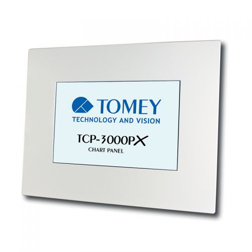
  • The Tomey chart panels offer the full range of tests for refraction. The TCP-3000PX has standard optotype tests, VA test, binocular tests (red-green), contrast sensitivity, colour vision and animation tests. The TCP-3000PX has a 24" display. When it comes to correction of associated heterophoria, the TCP-3000PX provides the complete and latest standard for a chart displaying monitor - this is because of its brilliant separation for right and left eye.

    TCP-3000PX Features:

    • Extensive polarised test chart selection
    • High quality LCD
    • Integrated computer
    • Multimedia tool
    • Colour vision test chart
    • Printable test reports
    • Programmable display sequences
    • Multilingual user interface
    • Large test field frame
    • Full MKH test sequence
    • 24" display
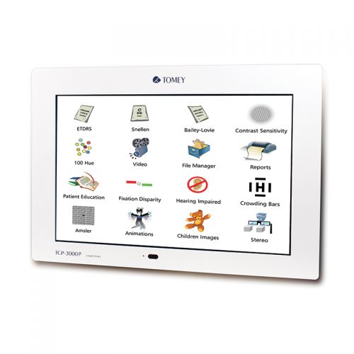
  • The Tomey chart panels offer the complete range of tests for refraction. The TCP-3000P has standard optotype tests, VA test, binocular tests (red-green), contrast sensitivity, colour vision and animation tests. The TCP-3000P has a 24" display and is a high quality all-rounder with extensive polarised test chart selection and integrated PC.

    TCP-3000P Features:

    • Extensive polarised test chart selection
    • High quality LCD
    • Integrated computer
    • Multimedia tool
    • Colour vision test chart
    • Printable test reports
    • Programmable display sequences
    • Multilingual user interface
    • 24" display
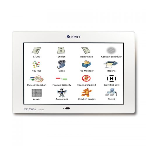
  • The Tomey chart panels offers the complete range of tests for refraction. The TCP-2000A has standard optotype tests, VA test, binocular tests (red-green), contrast sensitivity, colour vision and animation tests. The TCP-2000A has a 24" display and is Tomey's entry level chart monitor.

    TCP-2000A Features:

    • Programmable display sequences
    • Comprehensive test selection
    • Multimedia tool
    • Colour vision test charts
    • Contrast sensitivity test
    • Non-reflecting display
    • Individual test report function
    • 24" display
  • The Tomey CASIA2 is an outstanding machine, which fulfils the expectations of eye doctors specialised in all areas. CASIA2 impresses by intuitive and mostly automated handling, encouraged by an unbelievable measurement speed. Our software guides you easily from measuring through to analysis and then to the final report. Be inspired and see the eye from a different perspective. The CASIA2 has many impressive features need to be seen to be believed. Caught your interest? If so, you cam learn more about it by getting in touch with us.

    CASIA2 Features:

    • Testing application for Cataract / Glaucoma / Cornea Surgery
    • Glaucoma angle analysis (360)
    • Advanced imaging with high resolution and deep scanning depth (13mm)
    • Very fast scanning speed (50,000 A-scans/sec)
    • Corneal topography & IOL choice & calculation
    • Lens shape analysis & trend analysis
    • Phakic IOL simulation
    Download the Tomey CASIA2 Brochure here. Download the NEW CASIA2_User_Experience here.
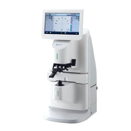
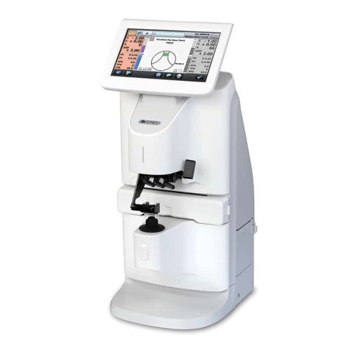
  • The Tomey TL-7000 uses a 117 point Hartmann sensor wave front technology meaning superior measurement accuracy and speed for all types of lenses is guaranteed. Green LED measurement light allows measure of high index lenses without the need of ABBE compensation. With the TL-7000 you are able to show your patients the transmittance of blue light (465nm) and of UV light (375nm) passing through their lenses simultaneously while measuring the optical power. Lens mark recognition support means various grid patterns will assist you to recognise the lens marks easier as well as to visualise the distortion of multifocal lenses. The grid patterns are changed by swiping the finger over the 7" touch panel. The TL-7000 also allows you to visualise the progression zone of multifocal lenses with the simple power mapping function. The use of PD-value and the L-value is recommended and is an exclusive function of the TL-7000.

    TL-7000 Features:

    • Wave front technology with Hartmann sensor (117 points)
    • Simultaneous measurement of UV/blue light and power
    • Lens mark recognition support
    • Simple power mapping
    • PD measurement
    • WiFi, LAN and RS-232C connection
  • The Tomey TL-6000 uses a 117 point Hartmann sensor wave front technology meaning superior measurement accuracy and speed for all types of lenses is guaranteed. Green LED measurement light allows measure of high index lenses without the need of ABBE compensation. With the TL-6000 you are able to show your patients the transmittance of blue light (465nm) and of UV light (375nm) passing through their lenses simultaneously while measuring the optical power. Lens mark recognition support means various grid patterns will assist you to recognise the lens marks easier as well as to visualise the distortion of multifocal lenses. The grid patterns are changed by swiping the finger over the 7" touch panel. The TL-6000 also allows you to visualise the progression zone of multifocal lenses with the simple power mapping function.

    TL-6000 Features:

    • Wave front technology with Hartmann sensor (117 points)
    • Simultaneous measurement of UV/blue light and power
    • Lens mark recognition support
    • Simple power mapping
    • LAN and RS-232C connection
  • EX-demo available

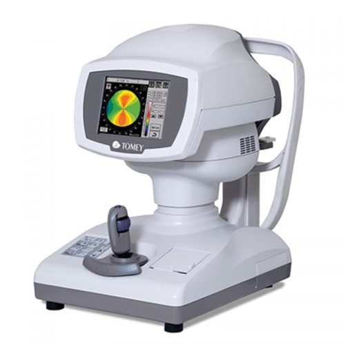
    The RT-7000 is a unique combination of topography, auto-refraction, keratometry and TSAS (tear stability analysis system) in one device. This multi-diagnostic instrument replaces four devices with all their functions. It is the perfect space and cost saving solution. Highly accurate measurements combined with short examination times and easy handling make working with the RT-7000 professional and quick. Alignment and measurement are done automatically - you just roughly align the system towards the patient's eye and the rest is taken care of by the instrument. The TSAS for analysing the tear film stability uses the light cone system and therefore offers you several measurement and analysis functions to detect patients with dry eye indication. For immediate understanding of the cornea topographic structure the RT-7000 implements the topographic indices KAI (Kerato-Asymetry Index) and KRI (Kerato-Regularity Index). These values are highlighted in colour (green = normal, yellow = suspect, red = abnormal) to provide you quick information about the corneal structure.

    RT-7000 Features:

    • 4 systems in 1 instrument
    • Colour touch screen
    • Different topography maps
    • Pupil & cornea measurement
    • Auto-alignment & auto-measurement
    • Tear stability analysis system (TSAS)
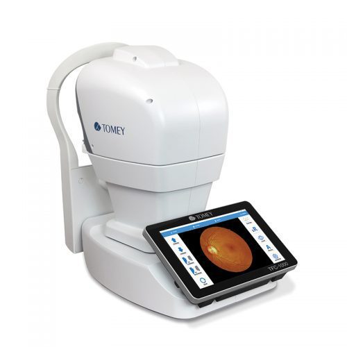
  • The TFC-1000 fully automatically generates a fundus image within 15 seconds using eye tracking and auto-focus as well as auto-measurement. The "KISS" (Keep It Short & Simple) principle had the highest priority in the software and workflow design. This makes the operation of the TFC-1000 extremely easy to handle. The user-friendly interface is very intuitive and provides the results in a very short time.

    TFC-1000 Features:

    • Automatic eye tracking
    • Non-mydriatic
    • Auto-focus & auto-measurement
    • Fundus image within 15 seconds
    • Multi-imaging display
    • Field of angle 45 x 45
    • 12 megapixel sensor
    • 9 fixation points
    • Anterior segment imaging
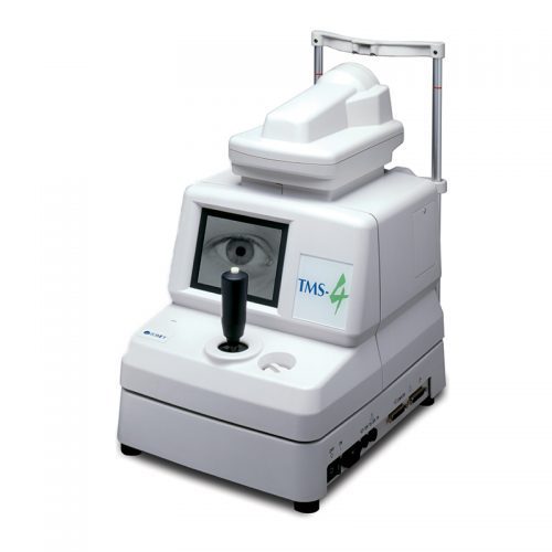
  • The TMS-4N guarantees high resolution with more than 60,000 data points. In addition the very short acquisition time (0.033 sec) assures you get the sharpest images for the highest accuracy. The TMS-4N has comprehensive software with single, dual and multiple map views allowing you to compare former data with new results. The Fourier analysis provides refractive information in 3mm and 6mm diameter range and also displays the spherical equivalent, regular astigmatism, asymmetry and higher order irregularity. Additional software applications such as Klyce statistics, keratoconus screening, enhanced height and height change maps are also available.

    TMS-4N Features:

    • Over 60,000 data points
    • Low light level cones
    • Auto-measurement & auto-select
    • Compatible software
    • Multilingual operation
    • Large patient database
    • Fourier refractive analysis
    • Keratoconus screening & other applications
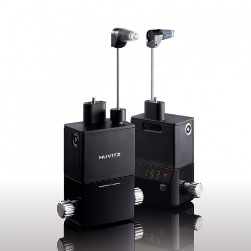
  • Using the Goldmann methodology for tonometry, the HT-5000 is Huvitz's applanation tonometer. It is mountable onto a variety of slit lamps and allows the user to determine approximate intraocular pressure.

    HT-5000 Features:

    • World standard Goldmann applanation tonometer
    • Digital display making it easy to read in dark environments
    • Wide grip dial for effortless adjustment of measuring prism
    • Optimised algorithm ensures the fast processing of IOP calculation
    • Instantaneous wake-up time from sleep mode
    • Powered by AAA batteries
    • Angled display matched to the angle of the user for comfortable observation
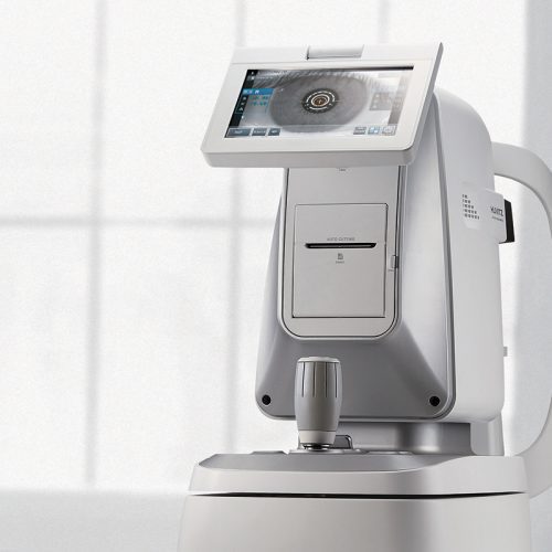
  • The Huvitz HRK-9000A combines everything into one simple device meaning patient workflow is improved by maximising efficiency.? It combines a auto-refractor keratometer, Contact Lens Fitting Guide (with colour camera), Meibomian Gland Analysis and Tear Film Break Up Time (TFBUT) functions in to a single instrument.  

    HRK-9000A Features

    • Subjective VA test
    • Wavefront technology
    • TFBUT measurement & Meibography
    • Wireless communication to other Huvitz products
    • Fully adjustable 7" touch colour display
    • Contrast sensitivity & glare test
    • Peripheral keratometry measurement
    • Auto-tracking feature
    • Colour View Mode: Full colour CCD camera and white LED source enables you to see eyes & contact lens fitting status previously only possible with slit lamps
    • Contact Lens Fitting Assistance Guide
    • Iris & Pupil diameter measurement
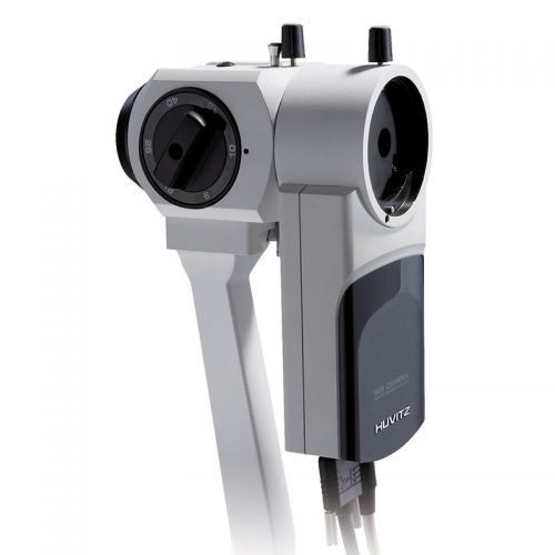
  • Huvitz Imaging System

    Enquire
    Enquire for Pricing
    Perfect solution to turn your HS-5000/5500 slit lamp into a complete image management system. Features:
    • A digital camera is attached to the binocular head where high resolution images can be captured and transferred to a pc.
    • Still images or videos are recorded using the joystick control on the base of the slit lamp.
    • Once the images are captured, they can easily be selected, manipulated and magnified to help you give an accurate diagnosis.
    • An MS Access Database allows you to search for symptoms, diagnosis information and record a patient's history, all within a user friendly environment.
    Camera and software included, please note that slit lamp must be bought separately.