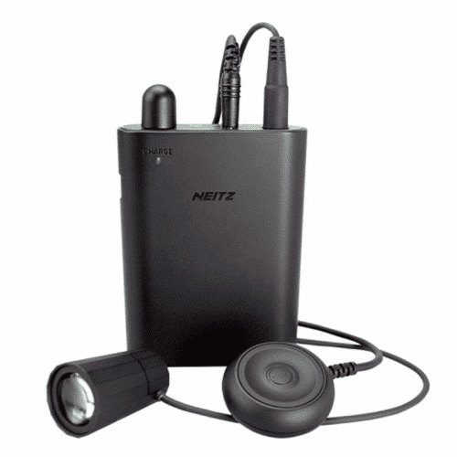
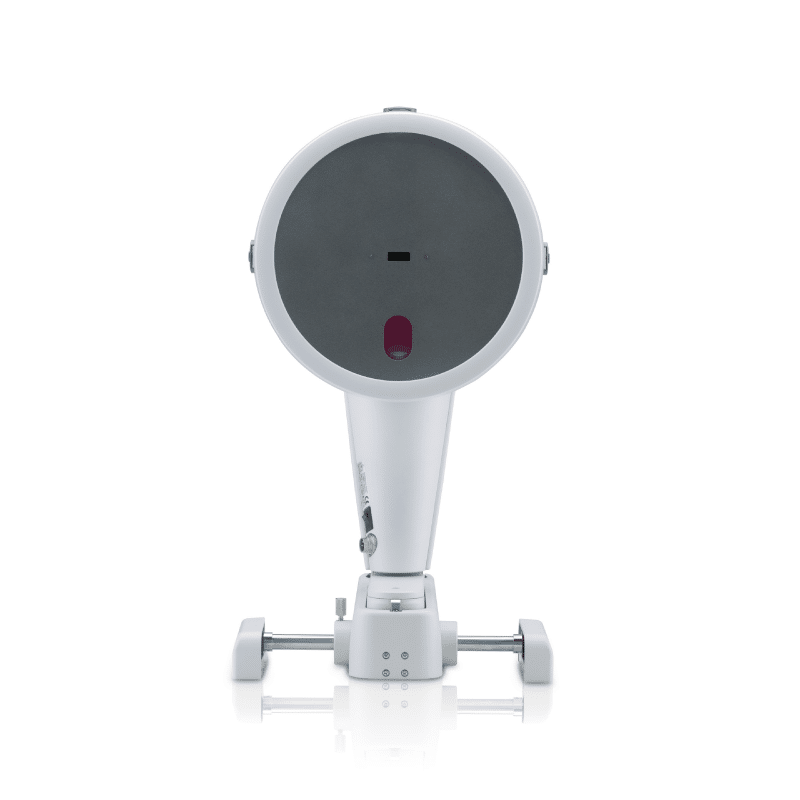
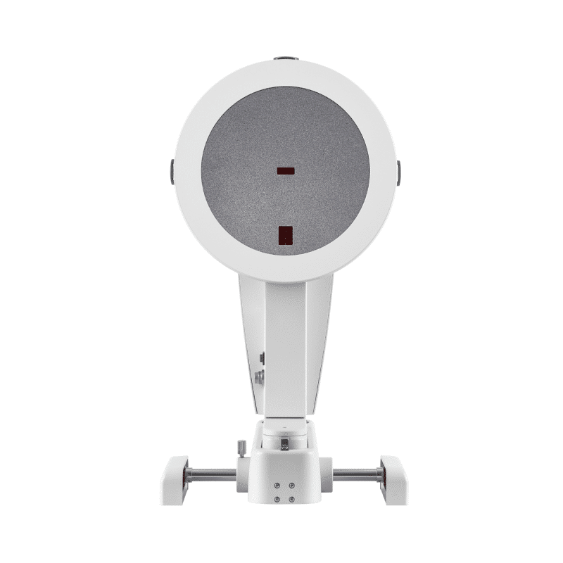
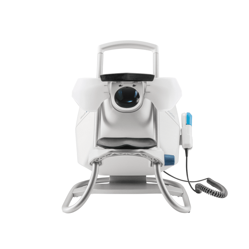
- World's highest illuminance; 32,000 lx for diameter of illumination field 100 mm, 38,000 lx for 80 mm.
- Small and lightweight, significantly reduced stress on the head and nose.
- Increased battery capacity, 2.5 hours of high-speed charging.
- Continuous illumination for approx. 3.5 hours at the maximum light intensity.
- Won the 2014 Good Design Award.
-
Sale!
-
Sale!
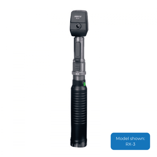
High quality product backed by ophthalmologists and certified orthoptists worldwide for accurate diagnosis - The Neitz retinoscopes use bulbs with precisely processed filaments of 0.05mm diameter, which create one of the sharpest streaks of light in the industry - Helps accurate diagnosis of the astigmatic axis - The beam can be turned 360 degrees easily - The anti-reflection filter provides a brighter and wider field of view - available in 3 model types
-
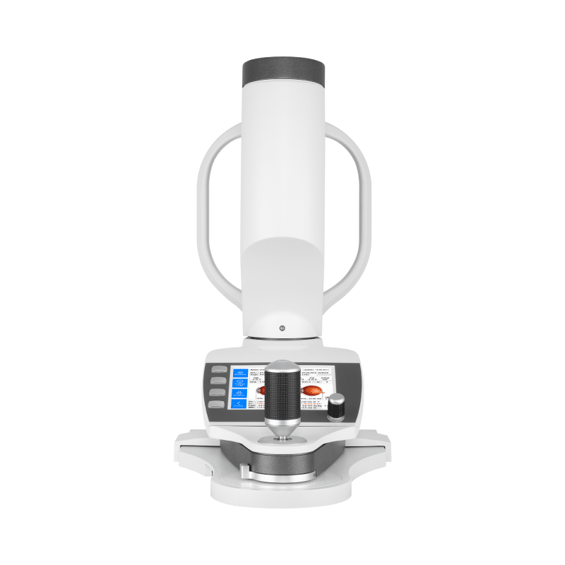
Evaluation of corneal biomechanical response, tonometry and pachymetry The revolutionary Corvis® ST records the reaction of the cornea to a defined air pulse with a newly developed high-speed Scheimpflug-camera that takes over 4 300 images per second. IOP and corneal can be measured with great precision on the basis of the Scheimpflug images.
- True IOP measurement with analysis of corneal hysteresis
- Link to Pentacam® for in depth ectasia analysis
- Progression analysis for CXL
- Biomechanical Tonometer and Corneal Analyser
-
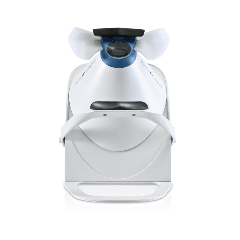
- Shorter examination times even for threshold tests
- No completely dark room required thanks to the closed construction
- Minimal footprint and maximal transportability
- Easy to service in the absence of moving parts
- Supra-threshold tests
- Advances test strategies, unique evaluation tools, efficient progression analysis
-
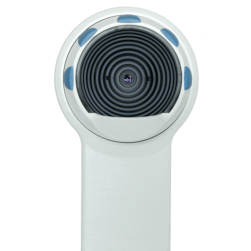
The Easygraph offers the ideal solution to practices struggling with limited space. Mounted directly on the slit lamp, this space-saving corneal topographer incorporates assessment of the cornea directly into the examination process. Equipped with proven measurement and device technology for contact lens fitting as well as precise and reliable diagnostics, the Easygraph has all it takes to be a real topographer.
*OPTIONAL Software extensions:
- Contact lens fitting: different contact lenses types and geometrics can be compared using fluorescein image simulation.
- Topographic Keratoconus Screening: eight different indices are calculated based on an analysis of the anterior corneal surface.
- Zernike Analysis: provides a means of describing irregularities of the cornea precisely.
-
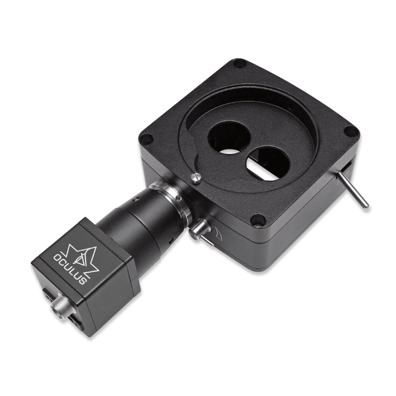
The ImageCam 3 - Universal Slitlamp imaging system offers high quality image and is one of the world smallest image documentation systems, it can be adapted to virtually all commercially available slit lamps.
- Avoid artefacts by taking images of the eye when it is at rest
- It varies the exposure time for you automatically
- Video recordings - create single images
-
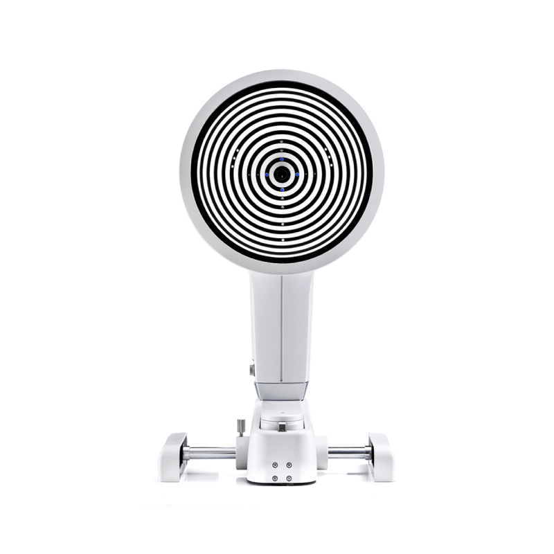
Multi-purpose Topographer Aside from Topography and Topographic keratoconus screening, the Keratograph 5M provides a large contact lens database and many analysis for daily practice. The built-in Keratometer and automatic measurement ensure the utmost accuracy and reproducibility. Detailed Display of the Cornea The software includes a reliable screening package for corneal detection, lens fitting and refractive surgery. Complete Documentation New Compare 4 Exams display. Changes from the first to the latest measurement can easily be displayed, reflecting the course of disease over time. JENVIS Pro Dry Eye Report Find the cause of dry eye syndrome quickly and reliably. Perform a comprehensive screening, using the measuring results as a basis for diagnosing dry eye syndrome. All results are documented in accordance with the Medical Products Law and summarised for your patient in a neat and easily understandable printout. Download the OCULUS - Keratograph 5M Brochure here.
-
All-in-one device for Myopia management
- Refraction
- Axial Length
- Keratometry
*OPTIONAL New GRAS Module
The Gullstrand Refractive Analysis System (GRAS) for short, is a refraction-analysis- module that is optionally available with the Myopia Master®. The GRAS examination compares individual measured parameters with the Gullstrand Eye and simulates the corresponding refractive changes in the spectacle plane. The Gullstrand parameters are replaced by the individual measurements of axial length, corneal radius and total refraction of the respective patient. OCULUS has adapted the Gullstrand eye to children. The advantage of the age-adapted GRAS module allows eye care professionals to detect pre-myopia. Download the Myopia Master® Brochure here. Download the sample Myopia Report here. -
The OCULUS Pentacam® AXL Wave is the first device to combine Scheimpflug tomography with Axial Length, Ocular Wavefront, Refraction and Retroillumination. Scheimpflug-based Tomography Pentacam® technology is the established gold standard, proven over many years. It measures, displays and analyses the Anterior Eye Segment, and is non-contact and tear-film-independent.
*Optional Software Package for Optometric Screening
- Belin/Ambrósio Display: Early detection of corneal irregularities and risk management for refractive surgery.
- Corneal Optical Densitometry: Objective analysis of corneal optical densitometry in different layers and zones.
- Show 2 Exams Topometric: For better comparison of two examinations results.
- 4 Maps Selectable: Customise your quad map, choose any of the available datasets to present on this screen.
*Optional Software Package Contact Lens
- Compare 4 Exams: objective and intuitive follow-up and documentation.
- Zernike Analysis: Zernike Analysis and determination of lower and higher order aberrations including normative data.
- Contact Lens Fitting: Integrated and expendable contact lens database and realistic fluorescein image simulation.
- CSP Report including CSP PRO: 250 Scheimpflug images covering a diameter of up to 18 mm are taken in the Sagittal height measuring process. All images of a Cornea Scleral Profile (CSP) scan are taken from the same visual axis without the need for eye movement.
-
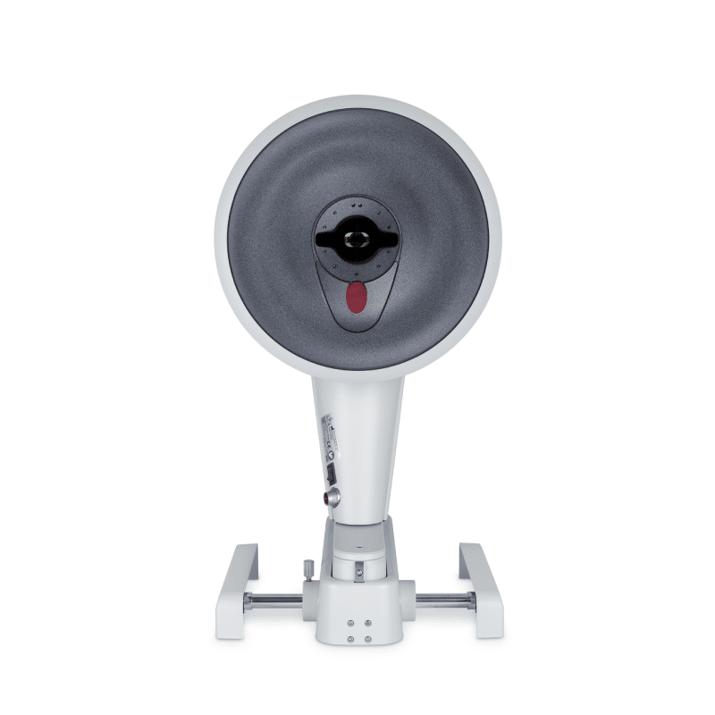
The Pentacam® HR supplies you with precise diagnostic data on the entire Anterior Eye Segment. The degree of corneal or crystalline lens density is made visible by the light scattering properties of the crystalline lens and is automatically quantified by the software. Measurement of the anterior and posterior corneal surfaces supplies the total refractive power as well as the thickness of the cornea over its entire area. The data on the posterior surface provide optimal assistance in the early detection of corneal changes. The rotating scan supplies a large number of data points in the centre of the cornea. A supplementary pupil camera captures eye movements during the examination for subsequent automatic correction of measured data. Equipped with intuitive and user-friendly software features to ensure patient safety.
*OPTIONAL Software Package Optometric Screening:
- Belin/Ambriósio Display: early detection of corneal irregularities and risk management for refractive surgery.
- Corneal Optical Densitometry: objective analysis of corneal optical densitometry in different layers and zones.
- Show 2 Exams Topometric: for better comparison of two examinations results.
- 4 Maps Selectable: customise your quad map, choose any of the available datasets to present on this screen.
*OPTIONAL More software options:
- CSP Report: Scleral Lens Fitting Guide
- 250 Scheimpflug images covering a diameter of up to 18 mm are taken in the measuring process. All images of a Cornea Scleral Profile (CSP) scan are taken from the same visual axis without the need for eye movement.
-
The OCULUS Pentacam® measures the entire anterior eye segment independent of tear film. Through high-resolution Scheimpflug images, the OCULUS Pentacam® calculates a motion-corrected 3D model of the anterior segment. It is also equipped with intuitive and user-friendly software features to ensure patient safety.
*OPTIONAL Software Package for Optometric Screening:
- Belin/Ambriósio Display: early detection of corneal irregularities and risk management for refractive surgery.
- Corneal Optical Densitometry: objective analysis of corneal optical densitometry in different layers and zones.
- Show 2 Exams Topometric: for better comparison of two examinations results.
- 4 Maps Selectable: customise your quad map, choose any of the available datasets to present on this screen.
*OPTIONAL More software options:
- CSP Report: Scleral Lens Fitting Guide
- 250 Scheimpflug images covering a diameter of up to 18 mm are taken in the measuring process. All images of a Cornea Scleral Profile (CSP) scan are taken from the same visual axis without the need for eye movement.
-
The modern device for Standard Automated Perimetry
- Short examination times even for threshold tests
- Advanced test strategies, unique evaluation tools
- Native Ethernet access
- Extended lifetime due to the absence of moving parts
- Small footprint and reduced weight for increased transportability
- No dark room required thanks to the closed design
- Practical carrying handle
- Height-adjustable measuring head


