Anterior Diagnostics

HomeAnterior Diagnostics
  • OCULUS – Easygraph

    Enquire
    Enquire for Pricing
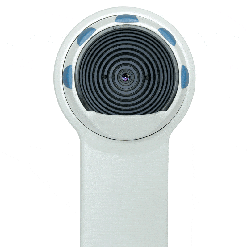
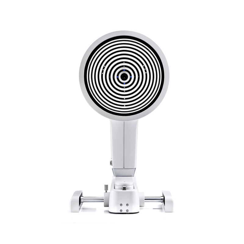
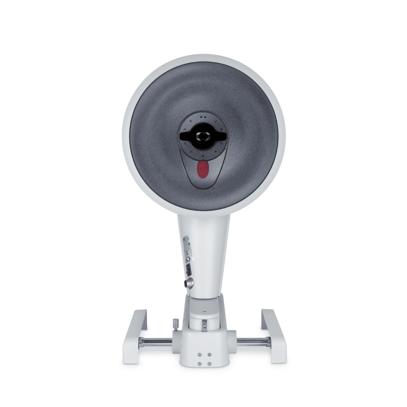
    The Easygraph offers the ideal solution to practices struggling with limited space. Mounted directly on the slit lamp, this space-saving corneal topographer incorporates assessment of the cornea directly into the examination process. Equipped with proven measurement and device technology for contact lens fitting as well as precise and reliable diagnostics, the Easygraph has all it takes to be a real topographer.

    *OPTIONAL Software extensions: 

    • Contact lens fitting: different contact lenses types and geometrics can be compared using fluorescein image simulation.
    • Topographic Keratoconus Screening: eight different indices are calculated based on an analysis of the anterior corneal surface.
    • Zernike Analysis: provides a means of describing irregularities of the cornea precisely.
    Download the Easygraph Brochure here.
  • Multi-purpose Topographer Aside from Topography and Topographic keratoconus screening, the Keratograph 5M provides a large contact lens database and many analysis for daily practice. The built-in Keratometer and automatic measurement ensure the utmost accuracy and reproducibility. Detailed Display of the Cornea The software includes a reliable screening package for corneal detection, lens fitting and refractive surgery. Complete Documentation  New Compare 4 Exams display. Changes from the first to the latest measurement can easily be displayed, reflecting the course of disease over time. JENVIS Pro Dry Eye Report Find the cause of dry eye syndrome quickly and reliably. Perform a comprehensive screening, using the measuring results as a basis for diagnosing dry eye syndrome. All results are documented in accordance with the Medical Products Law and summarised for your patient in a neat and easily understandable printout. Download the OCULUS - Keratograph 5M Brochure here.
  • The OCULUS Pentacam® measures the entire anterior eye segment independent of tear film. Through high-resolution Scheimpflug images, the OCULUS Pentacam® calculates a motion-corrected 3D model of the anterior segment. It is also equipped with intuitive and user-friendly software features to ensure patient safety.

    *OPTIONAL Software Package for Optometric Screening:

    • Belin/Ambriósio Display: early detection of corneal irregularities and risk management for refractive surgery.
    • Corneal Optical Densitometry: objective analysis of corneal optical densitometry in different layers and zones.
    • Show 2 Exams Topometric: for better comparison of two examinations results.
    • 4 Maps Selectable: customise your quad map, choose any of the available datasets to present on this screen.

    *OPTIONAL More software options:

    • CSP Report: Scleral Lens Fitting Guide
    • 250 Scheimpflug images covering a diameter of up to 18 mm are taken in the measuring process. All images of a Cornea Scleral Profile (CSP) scan are taken from the same visual axis without the need for eye movement.
    Download the OCULUS - Pentacam® Brochure here.  Download the OCULUS - Pentacam® Configuration Brochure here.
  • The Pentacam® HR supplies you with precise diagnostic data on the entire Anterior Eye Segment. The degree of corneal or crystalline lens density is made visible by the light scattering properties of the crystalline lens and is automatically quantified by the software. Measurement of the anterior and posterior corneal surfaces supplies the total refractive power as well as the thickness of the cornea over its entire area. The data on the posterior surface provide optimal assistance in the early detection of corneal changes. The rotating scan supplies a large number of data points in the centre of the cornea. A supplementary pupil camera captures eye movements during the examination for subsequent automatic correction of measured data. Equipped with intuitive and user-friendly software features to ensure patient safety.  

    *OPTIONAL Software Package Optometric Screening:

    • Belin/Ambriósio Display: early detection of corneal irregularities and risk management for refractive surgery.
    • Corneal Optical Densitometry: objective analysis of corneal optical densitometry in different layers and zones.
    • Show 2 Exams Topometric: for better comparison of two examinations results.
    • 4 Maps Selectable: customise your quad map, choose any of the available datasets to present on this screen.

    *OPTIONAL More software options:

    • CSP Report: Scleral Lens Fitting Guide
    • 250 Scheimpflug images covering a diameter of up to 18 mm are taken in the measuring process. All images of a Cornea Scleral Profile (CSP) scan are taken from the same visual axis without the need for eye movement.
    Download the OCULUS - Pentacam® Brochure here. Download the OCULUS - Pentacam® Configuration Brochure here.
  • The OCULUS Pentacam® AXL Wave is the first device to combine Scheimpflug tomography with Axial Length, Ocular Wavefront, Refraction and Retroillumination. Scheimpflug-based Tomography Pentacam® technology is the established gold standard, proven over many years. It measures, displays and analyses the Anterior Eye Segment, and is non-contact and tear-film-independent.

    *Optional Software Package for Optometric Screening

    • Belin/Ambrósio Display: Early detection of corneal irregularities and risk management for refractive surgery.
    • Corneal Optical Densitometry: Objective analysis of corneal optical densitometry in different layers and zones.
    • Show 2 Exams Topometric: For better comparison of two examinations results.
    • 4 Maps Selectable: Customise your quad map, choose any of the available datasets to present on this screen.

    *Optional Software Package Contact Lens

    • Compare 4 Exams: objective and intuitive follow-up and documentation.
    • Zernike Analysis: Zernike Analysis and determination of lower and higher order aberrations including normative data.
    • Contact Lens Fitting: Integrated and expendable contact lens database and realistic fluorescein image simulation.
    • CSP Report including CSP PRO: 250 Scheimpflug images covering a diameter of up to 18 mm are taken in the Sagittal height measuring process. All images of a Cornea Scleral Profile (CSP) scan are taken from the same visual axis without the need for eye movement.
    Download the OCULUS - Pentacam® AXL Wave Brochure here.
  • The Tomey CASIA2 is an outstanding machine, which fulfils the expectations of eye doctors specialised in all areas. CASIA2 impresses by intuitive and mostly automated handling, encouraged by an unbelievable measurement speed. Our software guides you easily from measuring through to analysis and then to the final report. Be inspired and see the eye from a different perspective. The CASIA2 has many impressive features need to be seen to be believed. Caught your interest? If so, you cam learn more about it by getting in touch with us.

    CASIA2 Features:

    • Testing application for Cataract / Glaucoma / Cornea Surgery
    • Glaucoma angle analysis (360)
    • Advanced imaging with high resolution and deep scanning depth (13mm)
    • Very fast scanning speed (50,000 A-scans/sec)
    • Corneal topography & IOL choice & calculation
    • Lens shape analysis & trend analysis
    • Phakic IOL simulation
    Download the Tomey CASIA2 Brochure here. Download the NEW CASIA2_User_Experience here.
  • EX-demo available

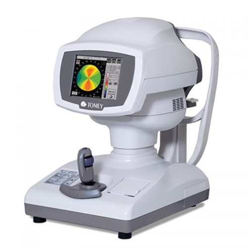
    The RT-7000 is a unique combination of topography, auto-refraction, keratometry and TSAS (tear stability analysis system) in one device. This multi-diagnostic instrument replaces four devices with all their functions. It is the perfect space and cost saving solution. Highly accurate measurements combined with short examination times and easy handling make working with the RT-7000 professional and quick. Alignment and measurement are done automatically - you just roughly align the system towards the patient's eye and the rest is taken care of by the instrument. The TSAS for analysing the tear film stability uses the light cone system and therefore offers you several measurement and analysis functions to detect patients with dry eye indication. For immediate understanding of the cornea topographic structure the RT-7000 implements the topographic indices KAI (Kerato-Asymetry Index) and KRI (Kerato-Regularity Index). These values are highlighted in colour (green = normal, yellow = suspect, red = abnormal) to provide you quick information about the corneal structure.

    RT-7000 Features:

    • 4 systems in 1 instrument
    • Colour touch screen
    • Different topography maps
    • Pupil & cornea measurement
    • Auto-alignment & auto-measurement
    • Tear stability analysis system (TSAS)
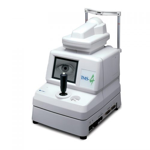
  • The TMS-4N guarantees high resolution with more than 60,000 data points. In addition the very short acquisition time (0.033 sec) assures you get the sharpest images for the highest accuracy. The TMS-4N has comprehensive software with single, dual and multiple map views allowing you to compare former data with new results. The Fourier analysis provides refractive information in 3mm and 6mm diameter range and also displays the spherical equivalent, regular astigmatism, asymmetry and higher order irregularity. Additional software applications such as Klyce statistics, keratoconus screening, enhanced height and height change maps are also available.

    TMS-4N Features:

    • Over 60,000 data points
    • Low light level cones
    • Auto-measurement & auto-select
    • Compatible software
    • Multilingual operation
    • Large patient database
    • Fourier refractive analysis
    • Keratoconus screening & other applications